© SAMS
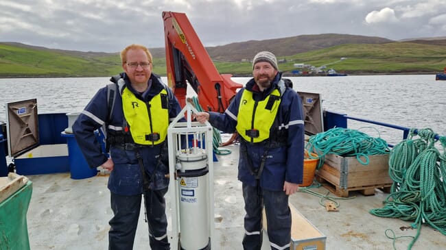
A high-tech underwater device that scans water samples for potentially dangerous algae blooms – the first of its type to be deployed in UK waters – has been operational for over a year at a Scottish Sea Farms site in Shetland.
The Imaging FlowCytobot (IFCB) uses a combination of lasers and cameras to detect, photograph, and identify phytoplankton species with the aim of alerting fish farm operators of potential harmful algal bloom (HAB) threats.
While phytoplankton are a critical part of the ocean ecosystem, some species can cause environmental issues when present in large numbers. Humans eating shellfish that have absorbed these toxic phytoplankton can become ill and blooms can also be fatal to farmed fish. Early warning of such phytoplankton blooms is therefore crucial to the aquaculture industry.
Since entering the water in spring 2023, the IFCB has photographed phytoplankton around the clock at 20-minute intervals and has already identified trends in the structure of the phytoplankton community from more than 38 million images. Thanks to funding from the Sustainable Aquaculture Innovation Centre (SAIC), researchers hope the observations made by the device will help them to better understand seasonal trends in harmful phytoplankton blooms.
The artificial intelligence developed for the IFCB marks significant progress in the world of phytoplankton taxonomy. Compared to traditional methods, which rely on the expertise of phytoplankton taxonomists and can take days of processing, the IFCB can provide near real-time information to farm-managers.
“It is notoriously difficult to predict when an algal bloom will occur, given the various environmental factors involved in its formation. The more warning we can give fish and shellfish farmers, the better the chance they have of mitigating the impact,” said Professor Keith Davidson, Scottish Association for Marine Sciences project leader, in a press release.
“With the IFCB, we have a virtual taxonomist on duty around the clock, identifying potential risks before a scientist has even looked down a microscope. It’s already showing us rapid changes over the courses of a day that we’ve never seen before. Traditional sampling methods use fixatives to preserve the sample for analysis but that can damage the cell. Being able to see live samples shows us the structure of the cell as it’s meant to be,” he explained.


