Hepatopancreatic microsporidiosis was first discovered in 2009 and is now endemic in many of the major shrimp farming countries, where it has affected the economic sustainability, production, profitability and supply of shrimp to global markets. It is caused by Enterocytozoon hepatopenaei (EHP) which is an infectious microsporidian parasite affecting hepatopancreas tubule cells of Penaeid shrimp species. Enterocytozoon hepatopenaei does not require other hosts for transmission, therefore it is a highly contagious disease through horizontal transmission. EHP infection is usually characterised by slow growth and a wide size distribution and can be positively correlated with high stocking densities (Geetha et al. 2022). It can be observed at the early stage of post-larvae production in hatcheries – primarily in Thailand, Vietnam, Indonesia, Malaysia and China. In a study by Geetha et al. (2022), EHP was shown to cause an average loss of US$813 per tonne of production in Indian Penaeus vannamei shrimp farming.
EHP infection in Pacific white shrimp
In a study by Kumar et al. (2022), the digestive enzymes, metabolism, physiology, immunity and growth responses of Pacific shrimp (P. vannamei) at different time intervals were investigated after 90 days of an EHP challenge and compared to a control group (not challenged).
The digestive enzymes α-amylase and lipase were significantly (P < 0.05) reduced in the EHP challenged group. The metabolic variables such as triglycerides (TG), total protein (TP), cholesterol (CL), glucose (GL), and alanine aminotransferase (ALT) were significantly (P < 0.05) low in the EHP challenged group. The non-specific immune response was affected by the EHP infection by decreasing the activity of alkaline phosphatase (ALP), catalase (CAT), γ- glutamyl transferase (GGT), total antioxidant capacity (T- AOC), superoxide anion (SOA), phenoloxidase (PO), and total hemocyte count (THC). The FCR of the challenged group was 3.01 ± 0.29, whereas the control was 1.64 ± 0.05. The growth of challenged animals was only 12.17 ± 0.80 g, while the control was 19.27 ± 0.5 g after 90 days of culture.
In another study by Subash et al. (2022), Penaeus vannamei shrimp were infected experimentally with EHP by injection of spores (~1x10⁵ spores/shrimp) and oral feeding infected hepatopancreas. There was a significant reduction in the immune parameters total haemocyte count (THC), catalase activity (CAT) and lysozyme activity (LYS) – at 6, 24 and 24 hours post-infection (HPI) respectively.
On the other hand, EHP infection resulted in significant higher levels of superoxide dismutase activity (SOD), prophenoloxidase activity (proPO) and respiratory burst activity (RBA) at 6 hpi. EHP infection resulted in higher oxidative stress (RBA) at 6 hpi and was counteracted by SOD and CAT to protect the host cell from oxidative damage. The Toll gene expression was activated as an early response, as EHP interacts with the host and activated the prophenoloxidase system and haemocytes migration to the infection site to fight the invaded EHP from 6 hpi. Although appropriate responses were triggered, it seems they are not efficient enough to eliminate the EHP. Also, the lysozymes that act from within the haemocytes seemed to be either deactivated by EHP or even maybe not released from the haemocytes, as the haemocytes fail to produce self-degranulation or are incapable of phagocytosing the intercellular parasites as the levels reduced from 24 hpi.

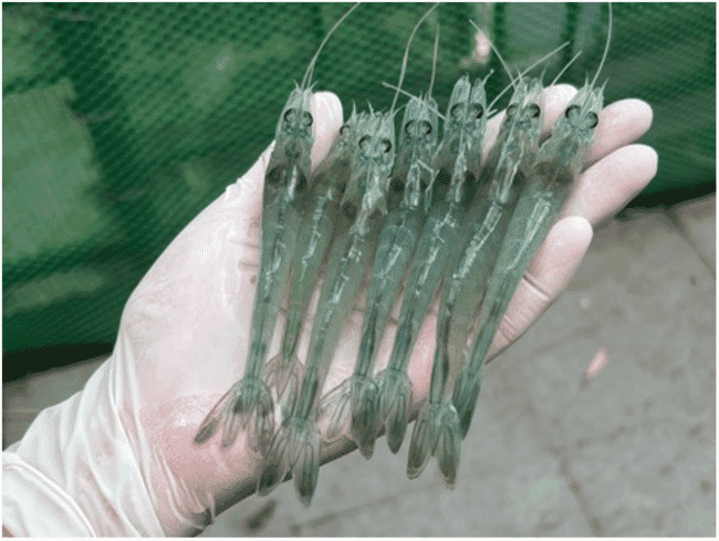
© Aranguren et al.
Cao et al. (2023) investigated the changes at the level of the transcriptome of the hepatopancreas after artificial challenge of healthy Penaeus vannamei with EHP. The differentially expressed genes (DEG) were closely related to immune response, and the enhancement of haematopoietic function and stimulation of Jak-STAT signalling pathway resulted in the ultimate activation of the immune system. The EHP infection increased lipid metabolism, and inhibited carbohydrate, amino acid metabolism and protein digestion – possibly the main causes affecting the normal growth of shrimp.
Haemocytin was reported for the first time in Litopenaeus vannamei (LvHCT) against EHP infection in a study by Sukonthamarn et al. (2023). Haemocytin is believed to be a major mediator of haemocyte aggregation and the prophenoloxidase (proPO) activation system. LvHCT suppression resulted in higher EHP copy numbers. LvHCT plays a vital role in shrimp innate immunity against EHP infection because the LvHCT gene transcript was upregulated after EHP infection and EHP copy numbers were increased in LvHCT-silenced shrimp.
Effect of water quality and culture system on EHP infection
In a study by Shen et al. (2019), slow-growth small shrimp, Penaeus vannamei, collected from earthen ponds showed a high prevalence of EHP infection (91.3 percent). Interestingly, the normal larger size shrimp in the earthen ponds also presented a high prevalence of EHP infection confirmed by first-step PCR (11 percent) and nested PCR (72.4 percent). The larger shrimp from a greenhouse had a lower EHP prevalence (10.6 percent) than normal larger shrimp from earthen ponds (72.4 percent), no EHP infection was detected by the first-step PCR in shrimp from the greenhouse. The authors suggested that shrimp cultures in greenhouses were less affected by EHP infection, as indicated by normal shrimp size and weight.
Six super-intensive ponds in Indonesia were evaluated for ammonia and nitrite levels, as well as EHP infection by Nkuba et al. (2021). A PCR test found two ponds were found to be positive for EHP, while nitrite and ammonia levels were above 1 ppm. The other four ponds were negative for EHP, and nitrite and ammonia levels were under 1 ppm.
In another study, it was observed that EHP infection can occur at a salinity of 2 ppt at a very low prevalence and severity, while the infection was higher in prevalence and severity at a salinity of 30 ppt (Aranguren et al. 2021).

EHP presence in other aquatic organisms present in shrimp ponds
In a study by Dewagan et al. (2023) at a shrimp farm in Maoming, China, shrimp and other organisms were investigated for EHP using PCR tests. EHP was detected in Litopenaeus vannamei, Penaeus monodon, crab, false mussel, and three dragonfly species (Anax parthenope, Pantala flavescens, and Ischnura senegalensis).
In the histopathological examination, EHP spores were found in naturally infected nymphs and adult dragonflies that were collected from the shrimp pond. Fluorescence in situ hybridisation results showed a positive signal for EHP infection in the body fat of dragonfly nymphs. Immature and mature microsporidian spores and late sporogonial plasmodium were observed in the cytoplasm of dragonfly nymphs using transmission electron microscopy.
The transmission of EHP from shrimp to dragonfly nymphs was confirmed via challenge experiments in which EHP-free dragonfly nymphs were cohabited with EHP-infected shrimp. The transmission of EHP from dragonfly nymphs to shrimp was demonstrated via the cohabitation of EHP-infected dragonfly nymphs with EHP-free shrimp and oral administration challenge experiments. The study confirmed that dragonflies can be another host for EHP, and that a horizontal transmission route of EHP is possible between dragonflies and shrimp.
In another study, in Malaysia, by Sajiri et al. (2023), EHP was detected in 82 aquatic specimens that belong to Arthropoda, Mollusca and Chordata phyla using the PCR targeting the genes encoding spore wall protein (SWP). The average prevalence of EHP by PCR was 82.93 percent for all three phyla (Arthropoda, Mollusca, and Chordata). These findings suggest the presence of EHP spores in aquatic organisms in the shrimp ponds which are potential transmission vectors (see table below).
The taxonomy of potential macrofauna-carriers collected from the shrimp ponds |
|||
Phyla |
Family |
Genus |
Description |
Arthropoda |
Varunidae |
Varuna, Metaplax, |
Family of crabs |
Sesarmidae |
Episesarma, Parasesarma |
Family of crabs |
|
Camptandriidae |
Paracleistostoma |
Family of crabs |
|
Nepidae |
Ranatra |
Family of water scorpions |
|
Mollusca |
Potamididae |
Mytella |
Family of bivalves |
Mytilidae |
Pirenella |
Family of bivalves |
|
Chordata |
Gobiidae |
Mugilogobius |
Family of bony fish |
Gut microbiota and EHP
Shen et al. (2021) investigated the link between EHP infection, growth retardation and intestinal microbiota in small, medium and large shrimp (P. vannamei). All the shrimp groups came from the same batch of post-larvae and were cultured in one pond under the same diet and environmental conditions. The small shrimp had the highest number of EHP copies. Denaturing gradient gel electrophoresis (DGGE) profiles showed that intestinal bacterial patterns of small and medium sized shrimp were similar, and different from the bacterial pattern in large shrimp. These results indicated that intestinal microbiota was influenced by EHP infection severity and the successive infection period of EHP. Another observation was that the relative abundance of Vibrio was highest in the small shrimp, suggesting higher susceptibility to bacterial invasion.

© Aranguren et al.
EHP and white faeces syndrome (WFS)
In an experimental study by Aranguren et al. (2021), WFS was reproduced successfully in P. vannamei. WFS is a disease of Penaeus monodon and P. vannamei associated with the presence of floating white faecal strings in grow-out ponds and clinical manifestations of white intestines and pale hepatopancreas. The pathogens responsible of this aetiology remain uncertain and it is suspected to be linked to a pathobiome caused by multiple enteric pathogens. The association of multiple pathogens is based on studies that WFS was observed in places that the prevalence and severity of EHP infection were high. In this study, an experimental reproduction of WFS in P. vannamei pre-infected with EHP was successfully accomplished after the challenge with a distinctive isolate of Vibrio parahaemolyticus. qPCR found that shrimp infected with both EHP and V. parahaemolyticus had a significantly higher load of EHP compared to shrimp infected with EHP alone. In this study, a synergistic relationship between EHP and V. parahaemolyticus isolate resulted in the manifestation of WFS.
Conclusions
EHP is a highly contagious disease known to have a devastating impact on shrimp production. Its geographical spread is increasing, so it is critical that shrimp producers have management protocols in place to prevent or mitigate the impacts of this disease.
There is no known effective treatment against EHP infection. But the impact of EHP spores and infection may be controlled by taking appropriate and effective actions (see below). Good biosecurity and an active monitoring programme are essential.
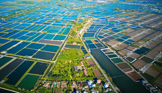
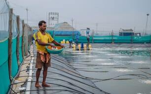
EHP has been widely recorded in shrimp farms in Thailand, Vietnam, Indonesia, Malaysia and China © Shutterstock
Key recommendations
- A biosecurity plan should be in place to prevent or mitigate EHP infections in farms.
- Only stock post-larvae that have been shown to be negative for EHP spores by PCR testing.
- Preparation of ponds before stocking with post-larvae free of EHP spores.
- The severity of EHP infection in low salinity (<10 ppt) ponds is significantly lower compared to higher salinity ponds (>25 ppt).
- The use of high-quality feed and functional feed additives that modulate the immune response and mitigate stress in shrimp to avoid secondary infections by vibriosis and white spot syndrome virus is recommended.
Sanitation
- Complete sanitation with quicklime and drying ponds for elimination of EHP spores.
- Sanitation of equipment and tools with potassium permanganate (KMnO4) (40 ppm for 15 minutes) (Aldama-Cano et al. 2018).
- Remove aquatic organisms that are potential passive or active carriers of EHP spores by filtration of new pond water.
- Good water quality and/or water parameters should be maintained throughout the production cycle.