The history of aquaculture in Iraq can be traced back to the mid to late 20th century, when the common carp Cyprinus carpio was introduced for the first time in 1955 into Al- Zaafaraniya fish farm at Baghdad city (Mhaisen, 1993). But in Kurdistan region, the culture industry developed only during the last twenty years or so, and now spread to many farms in the region (Coad, 2010).
C. carpio is the most widely frequently cultured fish. This species is characteristically hardy, high fertility, rapid growth, live on various types of foods, disease resistant, easy to reproduce and fast growing under proper conditions and live in a wider range of water temperatures 3-35° C (Gül et al., 2010).
Many fishes disasters in fish farms were caused by different considerable protozoa, which have direct life cycle and facilitate translocation from host to host making huge damages to fish wealth, i.e. Hines and Spira (1974) state that the parasite Ichthyophthirius multifiliis caused mortalities of common carp in hatcheries in North American.
The main objective of the present study was to know the ciliated protozoans that infect C. carpio from Ainkawa fish hatchery in Erbil city, Kurdistan region, Iraq.
Materials and Methods
A total of 210 fish belonging to common carp Cyprinus carpio were collected from Ainkawa Fish Hatchery, located northwest of Erbil city, Kurdistan region, Iraq. Fishes were collected by fishermen by using cast net and gill nets, during the period from August 2010 until the end of May 2011. Fishes were transported a live in a cool box with pond water to the laboratory of Parasitology, College Education/ Scientific Departments, University of Salahaddin. In the laboratory, fishes were identified after their capture according to Coad (2010).
The fishes were freshly examined by taking smears from skin, fins and buccal cavity through slight scraping. Gills were removed, put in a petri dish with water and macroscopically examined. Smears were also taken from the gills. All smears were examined under a light compound microscope at 40-100x magnification. Drawing of parasites was done by using a camera lucida. Olympus ocular micrometer was used to determine all necessary measurements for each parasite species.
Parasite identification was achieved by consulting some concerned taxonomical accounts (Bykhovskaya-Pavlovskaya et al., 1962; Hoffman, 1998).
Results and Discussion
A total of 210 common carp C. carpio were collected and inspected for parasites from Ainkawa fish hatchery. The present study showed the existence of six species of ciliated protozoans. The distribution of the parasites, their location on the fish host body, the prevalence and mean intensity of infection are summarized in Table (1). The following is an account on the description and measurements of these parasites:
Chilodonella cyprini (Moroff, 1902)
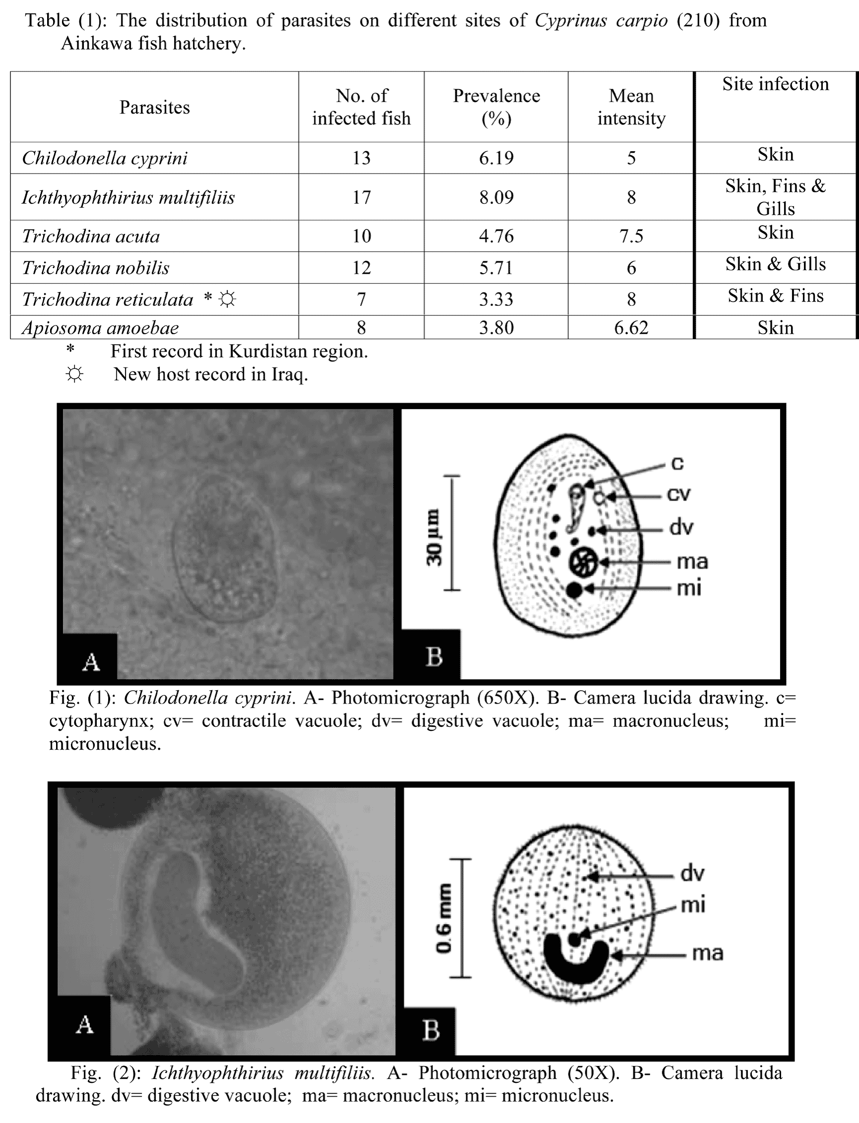
This parasite was found on the skin of C. carpio with a prevalence of 6.19% (Table 1). The body length 35-70 μm, width 20-35 μm. Body foliate, dorsoventraly depressed, dorsal side convex, ventral concave. Cilia present only ventrally in the form of several lateral parallel arched rows and large adoral band consisting of several larger cilia lying anteriorly to cytostome. Round macronucleus lies nearer to posterior end. Micronucleus lies adjacent to macronucleus. Two contractile vacuoles lie on diagonal line (Fig. 1).
The present specimens show a great similarity with the specimens of Ali et al. (1988), who reported it for the first time in Iraq on the skin and gills of Mystus pelusius from Tigris river in Baghdad. After that it was reported on C. carpio in Al-Zaafaraniya fish farm, Baghdad (Sadek, 1999). Also, it was reported in Kurdistan region on the skin of C. carpio from Ainkawa fish hatechery in Erbil city (Al-Marjan and Abdullah, 2009).
Ichthyophthirius multifiliis Fauquet, 1876
This ciliated protozoan was found on the skin, fins and gills of C. carpio with prevalence of 8.09% (Table 1). Body oval to round, 0.5-1.2 mm in diameter, ciliation uniform pellicle longitudinally striated. Horseshoe- shaped macronucleus visible without staining, micronucleus adhering to it in larger specimens (Fig. 2).
This parasite has been reported for the first time in Iraq from Mugil dussmien (Herzog, 1969). After that, it was reported from 23 different fish hosts including C. carpio (Mhaisen, 2012). In Kurdistan region, it was reported from different species from Darbandikhan lake, Lesser Zab river, Greater Zab river and Ainkawa fish hatchery (Abdullah, 2005; Abdullah and Mhaisen, 2006; Al-Marjan and Abdullah 2009).
Trichodina acuta Lom, 1961
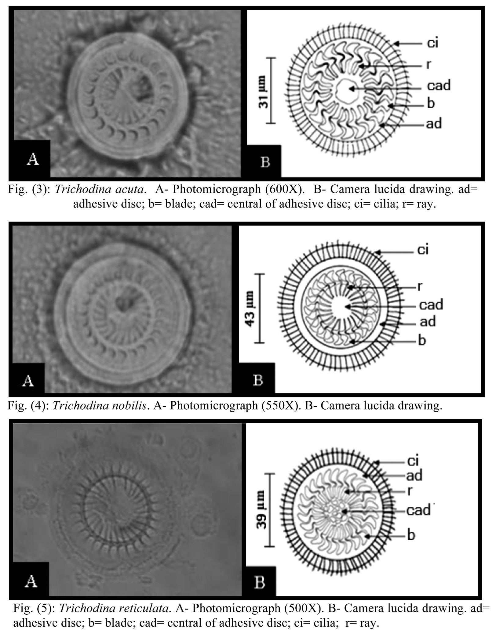
This ciliated protozoan was recorded on the skin of C. carpio with a prevalence of 4.76% (Table 1). This trichodinid with flattened and disc-shaped body falls within the range of the highest dimensions as a medium-sized species. Diameter of body 54-65 μm. Concave adhesive disc 44-55 μm in diameter surrounded by a finely striated distinct and broad band-like border membrane. The center of adhesive disc bears a clear area containing some darker spots, especially at periphery, and encircled by a heavily impregnated and wrinkled ring of central area. The denticulate ring composed of 20-23 denticles, 28-32
μm in diameter. The blade of denticle relatively short and blunt like a wide sickle and the ray of denticle thorn like. Central portion of blade broad, while ray of denticle thread like and pointed. Both blade and ray of denticle arranged at the same level. Length of blade 5-6 μm, length of ray 5-7 μm and length of span 13-15μm (Fig. 3).
The description and measurements of the present specimen are similar to those referred to by Al-Marjan and Abdullah (2007), which isolated from the skin, fins and gills of C. carpio from Ainkawa fish hatchery, Erbil province.
Trichodina nobilis Chen, 1963
This trichodinid was found on the skin and gills of C. carpio with a prevalence of 5.71% (Table 1). Large trichodinid 65-85 μm in diameter. Adhesive disc 55-74 μm in diameter surrounded by finely striated border membranes. Diameter of denticle ring 35-49 μm. The center of the adhesive disc without granule. Number of denticles 23-28. The blade of denticle narrowed at the base, while flattened at the apical, length of blade 7-10 μm. The ray thread like and pointed end, length of ray 8-13 μm. Both blade and ray arranged at the same level. Length of span 18-25 μm (Fig. 4).
This parasite was report for the first time in Iraq from the skin, fins and gills of C. carpio from Ainkawa fish hatchery, Erbil province (Al-Marjan and Abdullah, 2007).
Trichodina reticulata Hirschmann et Partsch, 1955
This species was isolated from the skin and fins of C. carpio with prevalence of 3.33% (Table 1). Medium to large discshaped trichodinid diameter of body 55-70 μm, adhesive disc 48-55 μm in diameter, denticle ring 30-38 μm. Number of denticles 22-26 denticles. Inner margin of denticle blade curved and roughly angular on outside margin, with squared distal end, length of blade 6-7 μm. Central part oblong, with oblong to rounded overlapping end. Projection of central part is invisible. Ray with little taper, tip blunt to square; length of ray 5-7 μm. Length of span 14-16 μm (Fig. 5).
This parasite was recorded for the first time in Iraq by Jori (2006) on the gills of Silurus triostegus from Al-Hammar marshes in Basrah city. No more hosts are known for this species in Iraq. So, C. carpio is now considered a new host for this ciliated protozoan in Iraq and the present study represents the first record of T. reticulata in Kurdistan region.
In addition to the three species of Trichodina mentioned above, 14 other species of this genus were reported in the Iraqi fishes namely: T. domerguei, T. nigra, T. cottidarum, T. gracilis, T. mulabilis, T. prowazeki, T. heterodentata, T. borealis, T. elegini and T. murmanica, T. ranae, T. pediculus, T. erbilensis and T. kurdistani (Mhaisen, 2012).
Apiosoma amoebae (Grenfell, 1887)
This parasite was obtained from the skin of C. carpio with a prevalence of 3.80% (Table 1). Peritrich with elongated conical body, with only anterior end contractile, size of body 18-65 X 15-30 μm. Macronucleus conical or round, micronucleus polymorphic. The body bears two rows of cilia one at the apical end, other at the mid of the body (Fig. 6).
This species was recorded for the first time in Iraq from Hypophthalmichthys molitrix in Babylon fish farm, near Hilla city (Ali et al., 1989). Later, it was recorded from C. carpio in Al-Zaafaraniya fish farm, Baghdad (Sadek, 1999). In Kurdistan region reported from C. carpio in Ainkawa fish hatchery in Erbil city (Al-Marjan and Abdullah, 2009).
According to Mhaisen (2012) five species of Apiosoma were known in different freshwater fishes in Iraq namely: A. amoeba, A. cylindriformis, A. minuta, A. piscicola and A. poteriformis.

September 2013


