First reports of red vent syndrome
These early reports from anglers, members of public and field based fishery scientists, were of occasional findings. However, during the summers of 2006 and 2007 there was a sudden increase in the number of observations of this condition.
The swollen vent condition soon gained the popular name of "Red Vent Syndrome" (RVS). RVS was recorded predominantly in adult, male and female one sea-winter fish, although a small number of two sea-winter salmon and sea trout (S. trutta) were also affected. No farmed salmon were reported with RVS.
Detailed laboratory investigations of salmon with swollen vents were first undertaken in November 2006. The Marine Laboratory in Aberdeen for Scotland and the Environment Agency and Cefas for England and Wales also began collating reports on the prevalence of red vents in returning fish from different rivers and catchment areas. By the end of 2007, most major salmon rivers in Scotland, England and Wales had confirmed records of affected fish.
The widespread nature of the observations and the significant public concern raised during the 2007 season prompted parallel studies in different institutions in an attempt to establish the cause and extent of the condition.
External observations of RVS
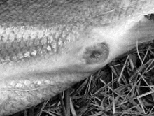
Affected fish showed different degrees of tissue damage around the vent and urogenital papilla, from mild swelling and reddening to severe protrusion, scale loss and haemorrhage.
In order to clarify and standardise reports, photographic guides including descriptions of mild, moderate and severe cases of RVS were developed in parallel by FRS for Scotland and the Environment Agency in England and Wales (example at http://www.frsscotland. gov.uk/FRS.Web/Uploads/Documents/R ed%20vent%20Scotweb.pdf ). Mildly affected salmon had a generalized reddening around the vent area and /or a number of small red spots (petechial haemorrhage) with little swelling of the vent area. Moderately affected fish also show a loss of integrity of the tegument observed through a breakdown of the epidermal layer and scale loss, while severely affected fish showed extensive loss of the epidermis and pronounced swelling with protrusion of the vent and bleeding.
Observations showed that the external lesions differed depending on how long fish had been present in freshwater. Some of the worst cases of RVS were recorded in estuarine caught salmon, while those captured after they had spent a period in freshwater showed stages of recovery. For example, early runners and summer fish, taken as broodstock and held in freshwater for 3 to 8 months, were examined at spawning time in hatcheries. They had either no or reduced reddening of the vent area and, in some cases, a full recovery of the skin was evident (Fig. 2).
The investigation into the cause

In order to improve understanding of the cause of RVS, tissue samples were taken and tested for a number of potential pathogens. Unaffected fish were also examined. Macroscopically, other than the vent area, fish showed good external condition and all internal organs appeared to be normal. The bacteriological, virological and molecular tests were negative for all known salmonid pathogens. However, moderate to large numbers of nematode parasites were found in the body cavity. In the affected fish, these nematodes were also in particularly high numbers within and around the vent region. The number of worms removed from this area could reach 100 individuals in a piece of tissue not bigger than 2.5 cm3. They were identified microscopically as a larval stage of the nematode Anisakis simplex sensu latum.
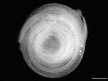
Anisakid nematodes are often found coiled, like a watch spring, measuring a few millimetres in diameter (Fig. 3), but when uncoiled larvae can measure up to 20 mm in length. They are normally seen encapsulated in a protective coat within the wall or external surfaces of the gut, pyloric caeca, liver and fat tissue. To the best of the authors' knowledge, these parasites have never been reported in the discrete region of the vent for any of the fish hosts this parasite infects.
Histological assessment

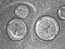
Histological sections of the vent area showed Anisakis larvae were embedded in the dermal, sub-dermal and muscle tissues surrounding the vent and uro-genital papilla. Changes observed included epidermal erosion, scale loss, moderate to severe dermatitis, haemorrhage and an inflammatory response dominated by eosinophilic granular cells (EGC), particularly in the estuary collected fish (Fig. 4). The EGC's were less prevalent or absent in fish caught later in the season, once they had spent some time in freshwater.
These tissue changes contributed to the external swelling and the general appearance of the inflamed vent. Considering affected fish showed a good general health status with no evidence of any other pathogens, the high number of nematode larvae observed within the vent area and the associated tissue reaction was postulated as the cause of the ‘red vent syndrome'.

Due to the novel location for the parasite, one of the first hypotheses that emerged was that the larvae in the vent could represent a sub species of the parasite. However, to date this hypothesis has not been tested and further research is being undertaken.
Discussion
Nematodes or "round worms" are common parasites in fish. About 11% of the over 750 genera of nematodes known for vertebrates can be found in fish hosts. Anisakis simplex is a commonly occurring nematode found in virtually all commercially important marine fish species in the Northeast Atlantic and, as a chronic infection, it does not result in mass mortality among its hosts.
The life cycle of Anisakis is very complex, involving marine mammals as the definitive final host, planktonic crustaceans as an intermediate host, and fish as transport hosts. In the North Atlantic ecosystem, the mechanisms that determine the occurrence and distribution of Anisakis are largely unknown. However, in these waters it appears to occur in fish from two ecologically different environments: the pelagic/oceanic and the benthic/coastal ecosystem. Salmon as a host act as an ‘accumulator', transporting the parasite from the intermediate host to the definitive host. The discrete region of the vent as a target tissue for the parasite represents a novel location so far only described for Atlantic salmon. The role and impact on the entire life cycle that the particular location (vent area) may have, is to be elucidated. Due to the consistent high numbers found in the area of all salmon analysed, we believe that the vent is unlikely to constitute an "accidental location".
Although the final natural host is a marine mammal, Anisakis may also affect humans when raw or undercooked fish is consumed, which raises a public health concern addressed by the Food Standards Agency.
From the studies performed between 2006 and 2008, the cause of the RVS in wild Atlantic salmon can be directly attributed to the presence of high numbers of Anisakis in the vent -urogenital papilla area. To date, there is no evidence that the condition has either led to mortality or prevented salmon from spawning in the past two seasons studied. Consequently, there is no basis to recommend that fish showing signs of RVS should be killed or removed from rivers.
Ongoing and future investigations
There is still much to learn about the cause and implications of RVS in wild Atlantic salmon. Monitoring for RVS has continued during 2008 with focus on determining the prevalence of the condition. Studies are also underway to assess the relationship between intensity of A. simplex and severity of vent lesions. Molecular investigations are also continuing to determine characteristics of the nematode as well as susceptibility of affected fish. These assessments are being undertaken at a range of selected sites throughout the UK.
Acknowledgements
The authors would like to acknowledge the assistance of the Association of Salmon Fishery Boards, fishery and riparian owners, biologists and fisheries officers throughout Scotland, England and Wales. The authors would also welcome notification of any future reports of RVS. Please contact the Fisheries Research Services in Scotland or the Environment Agency or Cefas in England and Wales.
June 2009


